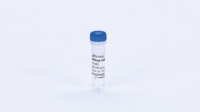
Cat. No. / ID: Y9240L
Features
- Non-competitive inhibitor of pancreatic-type ribonucleases e.g., RNases A, B and C
- Acidic, 52 kDa protein that is recombinantly expressed in E.coli carrying the porcine RNase Inhibitor gene
- Does not inhibit other RNases and modifying enzymes like polymerases and reverse transcriptases
Product Details
RNase Inhibitor is an acidic, 52 kDa protein that is a potent non-competitive inhibitor of pancreatic-type ribonucleases such as RNase A, RNase B and RNase C. The enzyme is provided as a fusion of the porcine RNase Inhibitor gene with a proprietary 22.5 kDa protein tag.
The enzyme is supplied in 20 mM Hepes-KOH, 50 mM KCl, 8 mM DTT, 50% glycerol, pH 7.5 at 25°C.
Performance
| Assay | Specification |
| Purity | >99% |
| Specific activity | 53,333 U/mg |
| Single-stranded exonuclease | 2000 U: <5.0% released |
| Double-stranded exonuclease | 2000 U: <1.0% released |
| Double-stranded endonuclease | 2000 U; no conversion |
| E. coli DNA contamination | 2000 U: <10 copies |
| RNase contamination | 2000 U: No detectable non-specific RNase |
Principle
The enzyme specifically inhibits RNases A, B and C. It inhibits RNases by binding noncovalently in a 1:1 ratio with high affinity. It does not interfere with the activities of other RNases and modifying enzymes like polymerases and reverse transcriptases.
Procedure
Instructions for using RNase Inhibitor are provided in the corresponding kit protocol in the resources below.
Quality Control
Unit activity was determined using the serial dilution method. Dilutions of the enzyme were made in 1X RNase Inhibitor Reaction Buffer and added to 1000 μL reactions containing 1mM cytidine 2’,3’-cyclic monophosphate, 1μg RNase A in a 1X reaction buffer containing 100mM Tris-Acetate, 1mM EDTA, pH 6.5. Absorbance at 286 nm was observed at 20-second intervals during a 5-minute reaction.
Protein concentration was determined using a 2.0 μL sample of enzyme analyzed at OD280 using a nanodrop ND-1000 spectrophotometer standardized using a 2.0 mg/mL BSA sample and blanked with product storage solution.
Physical purity was evaluated using 2.0 μL of concentrated enzyme solution loaded on a denaturing 4-20% Tris-Glycine SDS-PAGE gel flanked by a broad-range MW marker and 2.0 μL of a 1:100 dilution of the sample. Following electrophoresis, the gel was stained and the samples were compared to determine physical purity. The acceptance criteria for this test require that the aggregate mass of contaminant bands in the concentrated sample does not exceed the mass of the protein of interest band in the dilute sample, confirming greater than 99% purity of the concentrated sample.
Single-stranded exonuclease was determined in a 50 μL reaction containing 10,000 cpm of a radiolabeled single-stranded DNA substrate and 10 μL of enzyme solution incubated for 4 hours at 37°C.
Double-stranded exonuclease activity was determined in a 50 μL reaction containing 5,000 cpm of a radiolabeled double-stranded DNA substrate and 10 μL of enzyme solution incubated for 4 hours at 37ºC.
Double-stranded endonuclease activity was determined in a 50 μL reaction containing 0.5 μg of pBR322 DNA and 10 μL of enzyme solution incubated for 4 hours at 37°C.
E.coli contamination was evaluated using 5 μL samples of enzyme solution were denatured and screened in a TaqMan qPCR assay for the presence of contaminating E.coli genomic DNA using oligonucleotide primers corresponding to the 16S rRNA locus.
Applications
The enzyme is used for applications where the presence of RNases may pose a risk to RNA quality and experimental results:
- RNA isolation and purification
- cDNA synthesis
- RT-PCR and RT-qPCR
- In vitro transcription and translation
- RNase-free monoclonal antibody production

