Explore the biology, epidemiology and diagnostic options for common GI pathogens
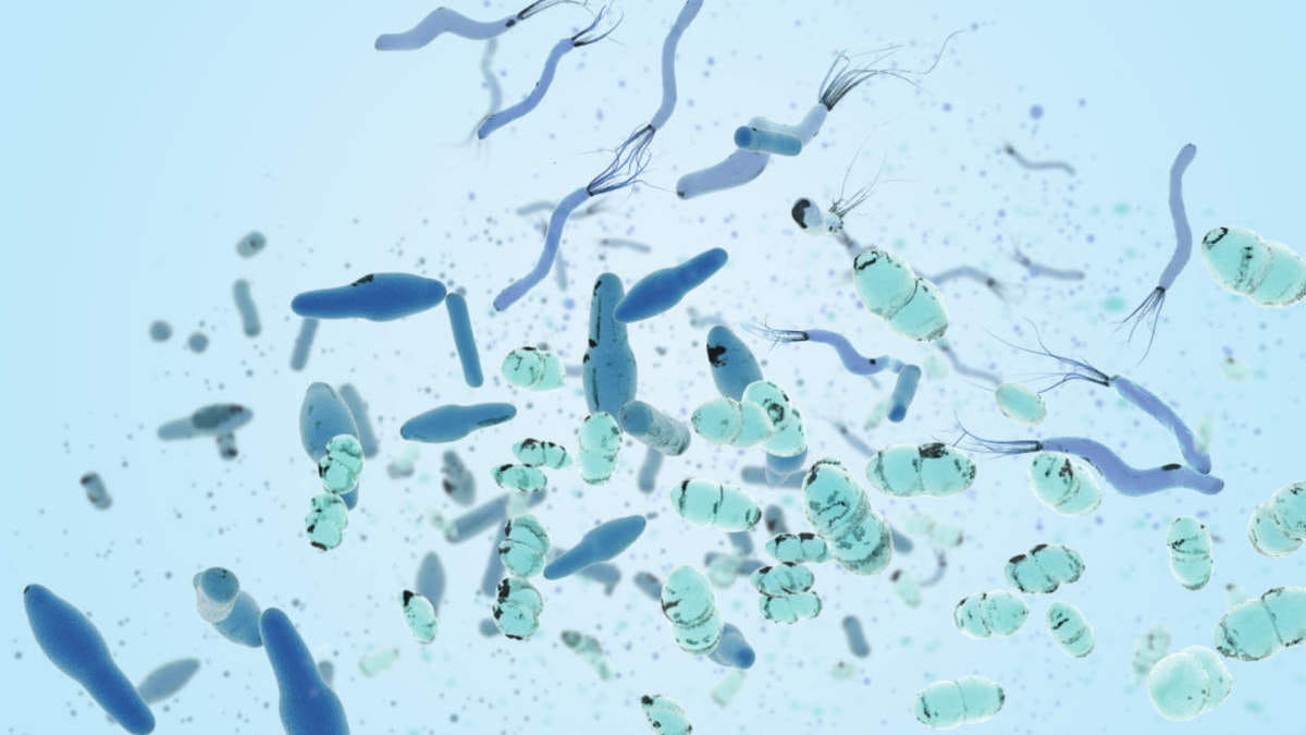
We all know a wide range of bacteria, viruses and parasites can cause gastrointestinal illness. But are you familiar with the major E. coli pathotypes linked to human disease and how they differ? Do you know the challenges involved in identifying many GI pathogens in the lab, including Entamoeba histolytica and Campylobacter spp? Welcome to our deep dive into GI pathogens. Read on to learn about some of the common pathogens that cause gastrointestinal infections.
Deep dive #1: E. coli pathotypes
Four important E. coli pathotypes
Not all pathogenic E. coli strains are the same. Some cause more severe disease than others, which is determined by distinct genetically-encoded virulence factors. There are five important E. coli pathotypes implicated in human disease. These can be distinguished from one another using molecular assays that detect their unique virulence factor genes.
1) EPEC
EPEC are classified into typical (tEPEC) and atypical (aEPEC) strains based on the presence of the adherence factor plasmid (pEAF). While both strains are important causes of diarrhea, tEPEC can cause more severe illness, leading to aggravation of malnutrition and persistent diarrhea.
EPEC’s key virulence factor is the eae gene. eae encodes a protein that allows the bacteria to adhere to human epithelial cells within the GI tract, causing destruction of cells.
In most cases, EPEC infections can be treated with hydration, although antibiotics are recommended for persistent infections or individuals at risk of severe infection (1).
2) ETEC
ETEC is the most common E. coli pathotype associated with diarrhea and is the leading cause of diarrhea among travelers and children. Its clinical symptoms range from mild diarrhea to a severe, cholera-like syndrome.
The bacteria is characterized by the production of heat-labile (LT) and heat-stable (ST) enterotoxins. These toxins are released after colonization in the small intestine and lead to watery diarrhea (2).
ETEC is usually self-limiting and can be treated with supportive care, including hydration. Antibiotic treatment can shorten the duration of diarrheal illness, but ETEC can be resistant to common antibiotics.
3) Shigella/EIEC
EIEC is an invasive strain of E. coli that is very closely related in virulence and other properties to Shigella. Both are characterized by profuse, sometimes bloody diarrhea, high fever and stomach cramps. Shigella causes an estimated 450,000 infections in the USA each year, with an estimated $93 million in direct medical costs (3).
Shigella/EIEC causes illness by penetrating epithelial cells and destroying the integrity of the epithelial lining (1).
While antibiotic treatment is recommended for severe shigellosis, drug resistant Shigella infections have been on the rise since 2016, with nearly 46% of infections being resistant in 2020 (4). Anti-diarrheal medications are not recommended, as these can worsen symptoms (1).
4) STEC
An estimated 265,000 STEC infections occur each year in the USA (5). Symptoms of STEC range from mild intestinal disease to hemorrhagic diarrhea. This can ultimately progress to a potentially fatal condition known as hemolytic uremic syndrome (HUS), especially in the very young and old. Approximately 5–10% of patients with STEC infections develop HUS.
The main virulence factors for STEC are the shiga toxins 1 (stx1) and 2 (stx2). STEC can carry either one or both toxin genes.
There are more than 400 serotypes of STEC, of which O157:H7 is the most common. The O157 serotype has resulted in large-scale outbreaks in several countries and is commonly associated with HUS (6). Identifying O157 and non-O157 E. coli serotypes is important for infection control and epidemiological surveillance.
Reducing the risk of HUS and severe GI disease

Deep dive #2: Identifying challenging GI pathogens
Many of the difficult-to-culture or difficult-to-identify organisms listed below can be easily missed in the lab using standard diagnostic methods. This is because the unique culturing conditions and tests needed to diagnose them are not routinely performed. More sensitive and reliable molecular assays like multiplex PCR may provide a better diagnostic yield for these and other GI pathogens. In fact, both Nebraska Medicine and the Mayo Clinic recommend multiplex PCR panel tests for certain patient populations (7,8).
Now, take a tour through some of the most challenging GI pathogens to identify in the lab.
Four GI pathogens that are especially difficult to work with and identify in the lab
| 1) Campylobacter spp. |
| 2) Shigella |
| 3) Salmonella spp. |
| 4) Intestinal parasites: Cyclospora cayetanensis, Cryptosporidium, Entamoeba histolytica, Giardia lamblia |


False-positive results
False positive results may be linked to sample contamination. They can have serious negative consequences for patients and healthcare institutions.
Patients may receive unnecessary or inappropriate changes in their treatment. For example, as discussed in deep dive #1, patients with a STEC infection should not be given antibiotics, as this can worsen their illness. As a result, a false-positive diagnosis could prove detrimental for these patients.
In addition, many GI pathogens are highly contagious and the presence of these illnesses can have ramifications for hospital function, including ward closure (19). False positives further run the risk of triggering public health alerts or investigations in the event that a pathogen may be foodborne or has human outbreak capacity.
False-negative results
False negative results can be caused by a low quantity of pathogen in the sample.
In many cases, individuals with GI illness will be placed on supportive or empiric treatment. However, if positive cases are not identified early, patients may not receive targeted treatment in a timely manner.
More to explore
References
- Hu J, Torres AG. Enteropathogenic Escherichia coli: foe or innocent bystander?. Clin Microbiol Infect. 2015;21(8):729-734. doi:10.1016/j.cmi.2015.01.015
- Kaper JB, Nataro JP, Mobley HL. Pathogenic Escherichia coli. Nat Rev Microbiol. 2004;2(2):123-140. doi:10.1038/nrmicro818
- Questions & Answers | Shigella – Shigellosis | CDC. https://www.cdc.gov/shigella/general-information.html
- CDC. COVID-19: U.S. Impact on Antimicrobial Resistance, Special Report 2022. Atlanta, GA: U.S. Department of Health and Human Services, CDC; 2022. https://www.cdc.gov/drugresistance/covid19.html
- Questions and Answers | E. coli | CDC. https://www.cdc.gov/ecoli/general/index.html
- Croxen MA, Law RJ, Scholz R, Keeney KM, Wlodarska M, Finlay BB. Recent advances in understanding enteric pathogenic Escherichia coli. Clin Microbiol Rev. 2013;26(4):822-880. doi:10.1128/CMR.00022-13
- GI Panel Guidance. Nebraska Medicine.
https://www.nebraskamed.com/sites/default/files/documents/for-providers/asp/GI_Panel_Guidance_10-18.pdf - Laboratory Testing For Infectious Causes of Diarrhea. Mayo Clinic Labs.
https://www.mayocliniclabs.com/en/-/media/it-mmfiles/Special%20Instructions/E/F/F/Laboratory_Testing_For_Infectious_Causes_of_Diarrhea - GBD 2016 Diarrhoeal Disease Collaborators. Estimates of the global, regional, and national morbidity, mortality, and aetiologies of diarrhoea in 195 countries: a systematic analysis for the Global Burden of Disease Study 2016. Lancet Infect Dis. 2018;18(11):1211-1228. doi:10.1016/S1473-3099(18)30362-1
- Janssen R, Krogfelt KA, Cawthraw SA, van Pelt W, Wagenaar JA, Owen RJ. Host-pathogen interactions in Campylobacter infections: the host perspective. Clin Microbiol Rev. 2008;21(3):505-518. doi:10.1128/CMR.00055-07
- M'ikanatha NM, Dettinger LA, Perry A, Rogers P, Reynolds SM, Nachamkin I. Culturing stool specimens for Campylobacter spp., Pennsylvania, USA. Emerg Infect Dis. 2012;18(3):484-487. doi:10.3201/eid1803.111266
- Fitzgerald C, Patrick M, Gonzalez A, et al. Multicenter Evaluation of Clinical Diagnostic Methods for Detection and Isolation of Campylobacter spp. from Stool. J Clin Microbiol. 2016;54(5):1209-1215. doi:10.1128/JCM.01925-15
- Vu DT, Sethabutr O, Von Seidlein L, et al. Detection of Shigella by a PCR assay targeting the ipaH gene suggests increased prevalence of shigellosis in Nha Trang, Vietnam. J Clin Microbiol. 2004;42(5):2031-2035. doi:10.1128/JCM.42.5.2031-2035.2004
- Kurtz JR, Goggins JA, McLachlan JB. Salmonella infection: Interplay between the bacteria and host immune system. Immunol Lett. 2017;190:42-50. doi:10.1016/j.imlet.2017.07.006
- Gal-Mor O, Boyle EC, Grassl GA. Same species, different diseases: how and why typhoidal and non-typhoidal Salmonella enterica serovars differ. Front Microbiol. 2014;5:391. Published 2014 Aug 4. doi:10.3389/fmicb.2014.00391
- Andrews JR, Ryan ET. Diagnostics for invasive Salmonella infections: Current challenges and future directions. Vaccine. 2015;33 Suppl 3(0 3):C8-C15. doi:10.1016/j.vaccine.2015.02.030
- Momčilović S, Cantacessi C, Arsić-Arsenijević V, Otranto D, Tasić-Otašević S. Rapid diagnosis of parasitic diseases: current scenario and future needs. Clin Microbiol Infect. 2019;25(3):290-309. doi:10.1016/j.cmi.2018.04.028
- CDC – Parasites.
www.cdc.gov/parasites - Norovirus | Guidelines Library | Infection Control | CDC.
https://www.cdc.gov/infectioncontrol/guidelines/norovirus/index.html