Getting to the Heart of miRNA Profiling
Micro RNAs (miRNAs) are small but mighty, playing a significant role in gene expression regulation. As key players in a multitude of biological processes, they are implicated in the pathogenesis of diseased conditions such as cell proliferation, differentiation, migration, apoptosis and inflammation. There’s mounting evidence of their potential as disease biomarkers, which has driven extraordinary developments in cancer, metabolic and disease research (1).
Navigating the challenges of miRNA profiling
Studying miRNA expression levels in biofluids such as serum, plasma, urine, etc., can provide an abundance of insights into the various disease mechanisms at play. However, miRNA profiling in biofluid samples is challenging because biofluids contain low RNA levels, high levels of inhibitors and are susceptible to many preanalytical variables. This has created a demand for dedicated solutions optimized for miRNA research – from high-quality miRNA extraction to efficient miRNA sequencing for miRNA expression insights.
Deciphering the role of miRNAs in disease research
Differential miRNA expression analysis is a key component of disease research. Two investigators at the Texas Heart Institute (THI), Lourdes Chacon-Alberty, M.D, Assistant Director of the THI Biorepository and Camila Hochman-Mendez, Ph.D., Assistant Director of Regenerative Medicine Research and Director of the THI Biorepository and Cell Profiling Lab, are providing novel insights into heart health through miRNA profiling. Ensuring consistency across replicates is critical when it comes to deciphering miRNA-seq data and drawing conclusions. Reliable miRNA solutions that can deliver highly consistent quality and results are therefore a prerequisite for research institutes such as the THI.
For almost 60 years, the THI has served as a premier nonprofit organization dedicated to reducing the devastating role of cardiovascular disease through innovative and progressive programs in research, education and improved patient care. “Through our translational basic and clinical research programs, we are learning more every day about the underlying causes of heart disease and the ways in which we can better treat and even prevent it”, explained Lourdes Chacon-Alberty. “As the leading cause of death in men and women worldwide, this is critical work.”
Differential miRNA expression in heart disease
Chacon-Alberty is one of six researchers within the Regenerative Medicine Research (RMR) team under the direction of Camila Hochman-Mendez at THI. One main focus of the RMR team is the role of differential miRNA expression in heart disease. Biorepositories, such as the one at the Texas Heart Institute, play a crucial role in the collection, storage, processing and distribution of biospecimens to support disease and biomarker research. From preserving the quality of the sample’s nucleic acids to minimizing sample-to-sample variation, biorepositories are indispensable for many research labs.
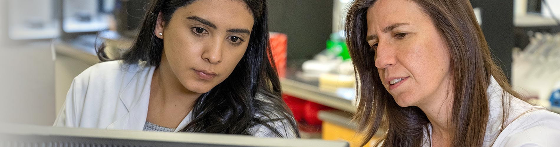
 Lourdes Chacon-Alberty is part of the Regenerative Medicine Research team at the THI Biorepository examining the significance of differential miRNA expression in heart disease.
 miRNAs from archived biofluids
THI’s archived biofluids serve as a rich and diverse source of miRNAs for gene expression studies conducted at the Institute. Additionally, the Biorepository & Cell Profiling Core (BRC) provides biobanking and standardized patient sample processing, profiling and storage services to academic and independent research institutions within the U.S. and Canada. It is home to the Cardiovascular Cell Therapy Research Network (CCTRN), the Cardiothoracic Surgical Trials Network’s (CTSN) samples.
 Camila Hochman-Mendez, Director of the THI Biorepository and Cell Profiling Lab, removes biorepository samples representing various tissue disease states from long-term cold storage.
 “From the biorepository, we typically use cryopreserved samples from de-identified patients that have been stored for more than 10 years. We have access to specific demographic/clinical characteristics of the patients. It is critical to ensure reliability within the replicates” Camila Hochman-Mendez emphasized. “In our investigations, we’ve also found it important to maintain consistency with the chemistries we use to isolate miRNA as well as to create miRNA libraries for next-generation sequence (NGS) analysis. Our lab has been using QIAGEN’s exoRNeasy Maxi Kit for exosome miRNA isolation from biofluids and the QIAseq miRNA Library Kit for the creation of our NGS miRNA libraries, both of which have provided highly consistent quality and results across the large variety of biorepository sample types we’ve used.”
When asked about some of their current projects, Lourdes Chacon-Alberty discussed two recent projects. “Currently, we’re in the process of analyzing miRNA expression in plasma-derived exosomes from lung transplanted patients. In our current study, we are focused on primary graft dysfunction (PGD), a severe type of lung injury occurring within the first 72h after lung transplantation. PGD is the most significant cause of morbidity and mortality after lung transplantation. We seek to identify differentially expressed miRNA in patients who develop PGD and those who do not. We have not published the results yet, however, we presented preliminary results in the International Society for Heart and Lung Transplantation Scientific meeting (ISHLT) at the end of April. Additionally, last year, we had the opportunity to publish an article in PLOS One in a collaborative project using bone marrow-derived cells (BMCs) cryopreserved cells from patients enrolled in two clinical trials conducted by NHLBI Cardiovascular Cell Therapy Research Network (CCTRN) - TIME (NCT00684021) and LateTIME (NCT00684060). Patients with ST-elevation myocardial infarction (STEMI) were treated with autologous bone marrow mononuclear cells (BM MNCs) harvested 3 and 7 days post-MI (TIME trial) or 2–3 weeks post-MI (LateTIME trial). In this paper, we showed that post-MI MNCs from these clinical trial patients, when harvested up to 3 weeks after MI, lack the therapeutic effects exhibited by healthy human BM MNCs when implanted into post-MI mouse hearts. We identified alterations in BM MNC composition and pro-inflammatory miRNA profile that may account for the BMC impairment post-MI.”
As evident from the research conducted in THI, miRNA profiling is driving advances in cardiology research (2). By exploring differential gene expression, researchers can unravel novel insights into the complex disease mechanisms at play, leading to developments in disease treatments and therapeutics.
Click here to learn more about QIAGEN miRNA NGS technologies.
Click here to learn about QIAGEN Genomic Services’ offering for miRNA-seq.
The views and opinions are those of the Investigators, and not necessarily those of Texas Heart Institute. Texas Heart Institute receives no remuneration and makes no endorsements of any kind nor of any product or company.Meet the researchers
We interviewed both Lourdes Chacon-Alberty and Camila Hochman-Mendez about their background, what led them to become scientists and what drives their interest in heart disease and research. Here’s what they had to say:
Lourdes Chacon Alberty

Science and math were always my favorites classes since I was a little girl. I remember when I was in high school, I used to help my teacher to explain it to my classmates. There are many things that influenced my interest in research, one of them is that I’m naturally curious about scientific phenomena, and I was inspired by people who strive to make a difference in the lives of others. In addition, I had great mentors and teachers who gave me the knowledge, tools and support to achieve my dreams.
To be a scientist requires commitment and passion because is a long learning process and it never stops because science is always evolving. Despite the hurdles, being a scientist remains an incredibly rewarding career path. There is no greater privilege than the potential to positively improve people’s lives and to help to solve global problems. It brings tremendous meaning and joy to the day’s work. In addition, working in a team environment in a lab can be a lot of fun, especially when discoveries are being made. The moment I started working in the lab, I realized this is what I always, always wanted to do.
I graduated from the Catholic University of Honduras with a medical degree. I then completed a master’s in clinical translational management at the University of St. Thomas, Houston, Texas. This program focused on bringing innovations from the bench to the bedside. Currently, I am a research scientist and assistant director of THI Biorepository Core at The Texas Heart Institute. I have been a member of the Texas Heart Institute team for 4 years. Here, at Texas Heart Institute, we are very fortunate to have access to many resources – a clinical research center, an editorial team, a dedicated grant writer, cardiovascular seminars and conferences, shared equipment. Additionally, we are located in the Texas Medical Center that allows us to make great collaborations.
Camila Hochman-Mendez

References
- Condrat et L. (2020) miRNAs as biomarkers in disease: Latest findings regarding their role in diagnosis and prognosis. Cells. 9(2): 276.
- Wang et al. (2020) Impaired therapeutic efficacy of bone marrow cells from post-myocardial infarction patients in the TIME and LateTIME clinical trials. PLoS ONE 15(8):e0237401.https://doi.org/10.1371/journal.pone.0237401