Bone regeneration is a complex physiological process, critical to both the healing of fractures and continuous bone remodeling in adults. Sometimes, a large amount of bone regeneration is required, and the normal process is inadequate. This can happen in cases of trauma, infection, tumor resection or skeletal abnormalities like osteoporosis (1).
A number of regenerative medicine strategies including autologous bone graft, allograft implantation or use of growth factors can be used to treat these cases. However, new, alternative approaches to support skeletal muscle and bone regeneration remain an important medical need.
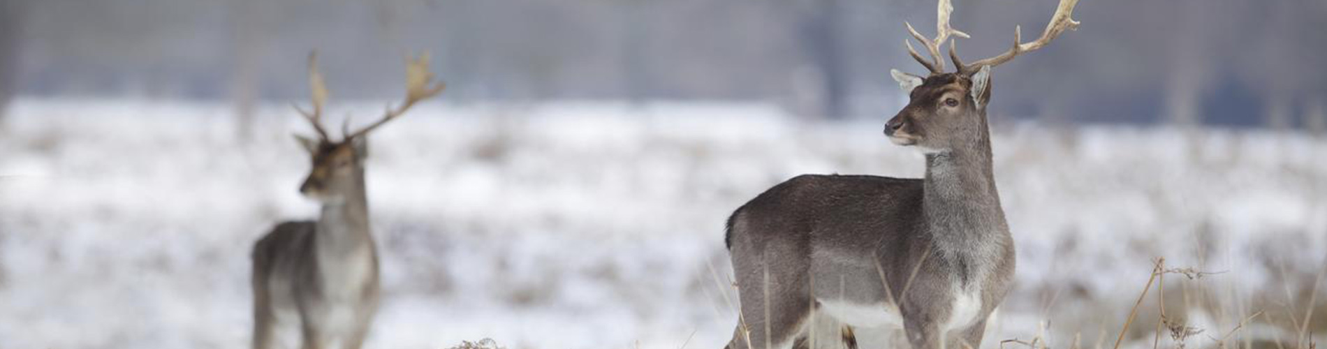
Dr. Dai Fei Elmer Ker and Dr. Peter Yang from the Stanford University School of Medicine, along with their colleagues, have been investigating rapid bone regeneration in quite a unique model – deer antlers! They recently published their findings in this article (2) in the journal Stem Cell Research & Therapy.
The inspiration to study bone regeneration in deer antlers came to Dr. Yang during a vacation in Canada while hearing about wild deer fun facts from a tour guide. Particularly striking was the fact that deer antlers can grow up to 2 cm in only one day. It would require an entire year for human femur bone to grow that much during puberty (3, 4).
The researchers established an in vitro model comparing deer antler-derived reserve mesenchyme (RM) cells with human mesenchymal stem cells – a clinically promising therapeutic target for cell-based regenerative medicine (5). They performed comparative RNA sequencing using RNA isolated with the RNeasy Plus Mini Kit and analyzed their data using several types of bioinformatics software, including Ingenuity Pathway Analysis (2).
The team identified gene candidates based on these criteria: differentially expressed genes upregulated more than five-fold in control versus treatment conditions; or genes that were uniquely expressed in the fallow deer RM cell dataset. From this list, the deer genes uhrf1 and s100a10 were identified as genes of particular interest.
Subsequent immunofluorescence studies showed expression of uhrf1 and s100a10 in regenerating deer antlers. Gene overexpression and knockdown studies demonstrated the proliferation contributions of uhrf1 and mineralization capabilities of s100a10 (2). Both uhrf1 and s100a10 also appear to be linked to human bone development. In summary, these findings might bring exciting innovations for treating human bone fractures and diseases in the future (3).
Are you using NGS for your studies? Explore QIAGEN’s complete automated NGS workflow solutions by downloading the e-book!
References:
1. Dimitriou, R. et al. (2011) Bone regeneration: current concepts and future directions. BMC Med, 9:66. Link
2. Ker D.F.E. et al. (2018) Identifying deer antler uhrf1 proliferation and s100a10 mineralization genes using comparative RNA-seq. Stem Cell Res Ther, 9(1):292. Link
3. RNA-seq blog https://www.rna-seqblog.com/genes-behind-rapid-deer-antler-growth-hardening-identified-through-rna-sequencing/
4. Sissons, H.A. and Kember, N.F. (1977) Longitudinal bone growth of the human femur. Postgrad Med J, 53(622):433–7. Link
5. Mendicino, M. et al. (2014) MSC-based product characterization for clinical trials: an FDA perspective. Cell Stem Cell. 14:141–5. Link
